CBCT Dental Tomography - Poznan
What is CBCT computer tomography?
Computer tomography (CBCT, 3D imaging, cone beam tomography) is a procedure that is becoming the standard in modern dental treatment. It is a test that presents the tissues of the oral cavity in a three-dimensional view. Importantly, it is the only image where both soft tissues and bone tissue are visible. We get a multi-plane image of the jaw and mandible, which can be viewed in many sections.
CBCT tomography - like classic tomography - uses X-ray radiation (RTG). The difference lies in the type of radiation beam. In CBCT cone beam tomography, the beam takes the shape of a cone, hence its colloquial name. We get a three-dimensional image, which allows for precise planning of dental treatment.
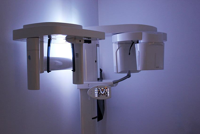
Computer tomography is currently widely used in all branches of medicine. In our Clinic in Poznań, we perform this examination using high-class Orthophos SL 3D equipment, which allows for the performance of a CBCT examination, which is a faster and safer variant of classic computer tomography, created specifically for the needs of dentistry. The CBCT tomography device emits a smaller dose of X-ray radiation compared to imaging with classic tomography.
What is a 3D image useful for?
The effectiveness of treatment largely depends on the diagnostic procedure.
3D examination is extremely useful during patient diagnostics, as it replaces a number of other X-ray images, including:
- pantomographic
- cephalometric
- wing-bite
- temporomandibular joint
- paranasal sinuses.
During the CBCT examination, the doctor assesses:
- the amount and condition of bone tissue
- the exact location of nerves and blood vessels
- maxillary sinuses
- tooth roots
- the presence of inflammatory conditions in the structures of the jaw and mandible
Thanks to this, the doctor is able to plan the course of the surgical or implantological procedure very precisely.
In the case of planned implant placement, the dentist in a special program designs their position with accuracy to tenths of a millimeter.
In the case of endodontic (root canal) treatment, CBCT computed tomography allows for precise estimation of the length of root canals, assessment of their condition and location of additional, atypically located canals.
CBCT examination is used in every field of dentistry. It is most often used in the diagnosis of:
- temporomandibular joint disorders,
- dental diseases and disorders,
- malocclusion,
- the degree of caries progression,
- periodontal diseases and bone defects of the jaw or mandible
When should a CBCT be performed?
The CBCT examination is ordered by the dentist during the consultation, according to the current diagnostic needs. The examination is performed on-site and "on the spot" at our Clinic in Poznań. The image of the jaw and/or mandible is obtained almost instantly, allowing the doctor to immediately discuss the result with the patient.
To perform a 3D image, it is necessary to remove jewelry, glasses, and take out any removable dentures or orthodontic appliances from the mouth. Dental tomography is performed in a standing or sitting position. During the examination, you should not move. The device emits X-ray radiation and its detector rotates 360°. The images are sent to a computer, where they are processed by an algorithm. The final result is a three-dimensional image of the analyzed structures.
Is this examination safe?
The examination itself is completely non-invasive and painless for the Patient. The examination time is only a few seconds. The amount of X-ray radiation is reduced to a minimum thanks to the use of digital technology and dental CBCT tomography instead of traditional computer tomography (tenfold reduction in radiation).
The smallest possible area of the oral cavity is always scanned, and during the examination, the Patient is protected with a lead apron.
Contraindications for performing CBCT examination
The main contraindication for the examination is the patient's pregnancy. Before proceeding with the imaging, the doctor should be informed about the pregnancy.
Images using CBCT tomography in children are only taken when it is necessary for further treatment.
Dental X-ray - Poznan
What is a dental X-ray?
An X-ray of a tooth - also commonly referred to as a radiograph - is an examination that uses X-ray radiation to gather information about the condition of the teeth before starting treatment. In dentistry, both point, panoramic (pantomographic), and cephalometric X-rays are used. The type of X-ray is chosen depending on the needs.
Point X-ray - It is used to assess the condition of the tooth or the advancement of the inflammatory process. It is also performed to check the filling of the canals or the effectiveness of the treatment.
Panoramic X-ray - widely used in dental diagnostics to assess the condition of the entire oral cavity, as all teeth are visible in the image. The pantogram allows to determine which teeth require urgent dental intervention, whether and how wisdom teeth affect the overall dentition, and is extremely helpful in orthodontic treatment.
Cephalometric X-ray - used to assess malocclusions and their impact on surrounding tissues. The image also includes the bones of the entire skull and sinuses in a lateral view.
When should an X-ray examination be performed?
An X-ray is most often taken before planned root canal treatment, tooth extraction, or the creation of a new prosthesis. It is also used to diagnose pathological changes at the roots, to detect the presence of caries and to identify impacted teeth. The X-ray image also allows to confirm the presence of cysts or the existence of tumorous changes within the facial skull.
Is an X-ray safe?
Diagnostic imaging using X-rays is safe for the patient. The dose is so small that it does not affect their health.
It is not recommended to perform X-ray imaging on pregnant women and it should be limited in children under 5 years of age.
Price List
For imaging studies, we use the ultra-modern SIRONA - Orthophos SL 3D tomograph
|
Digital panoramic image (pantomogram) 100 zł
The so-called "panoramic" examination complements the extraoral dental examination ("check-up"). Thanks to it, the dentist can see what is invisible to the naked eye - inflammation in the bone, decay under the filling, or impacted teeth. According to global standards, it should be performed every two years. |
|
Digital Cephalometric X-ray 100 zł
It illustrates the facial skull in a lateral view. It shows the way of biting and the mutual arrangement of the mandible and maxilla. Helpful in the diagnosis of malocclusion and orthodontic treatment. |
|
Tomography (CBCT) 180 - 320 zł
The standard in modern dental treatment - it illustrates the bone in three dimensions, allowing you to "see it from every side". CBCT is a dental variant of classic computer tomography with a significantly reduced dose of radiation. In our Clinic, we use the ultra-modern SIRONA SL 3D tomograph.
CBCT one area 180 zł
CBCT 1 arch 220 zł
CBCT 2 arches 320 zł
|